
At the Clinic
At the Clinic
The peripheral nerve injury clinic brings together a team of healthcare specialists who play different roles in your care.
At your appointment, you may see a few of these specialists depending on your specific concerns and where you are in your recovery.
You might also be asked to complete diagnostic tests or functional assessments to better understand your injury, functional changes, or monitor your recovery progress.
Your Care Team
Your care team is made up of healthcare professionals with a wide range of skill sets and expertise. This section provides information on:
- What their role is in your care.
- What kind of information they can provide you with.
Physiatrists
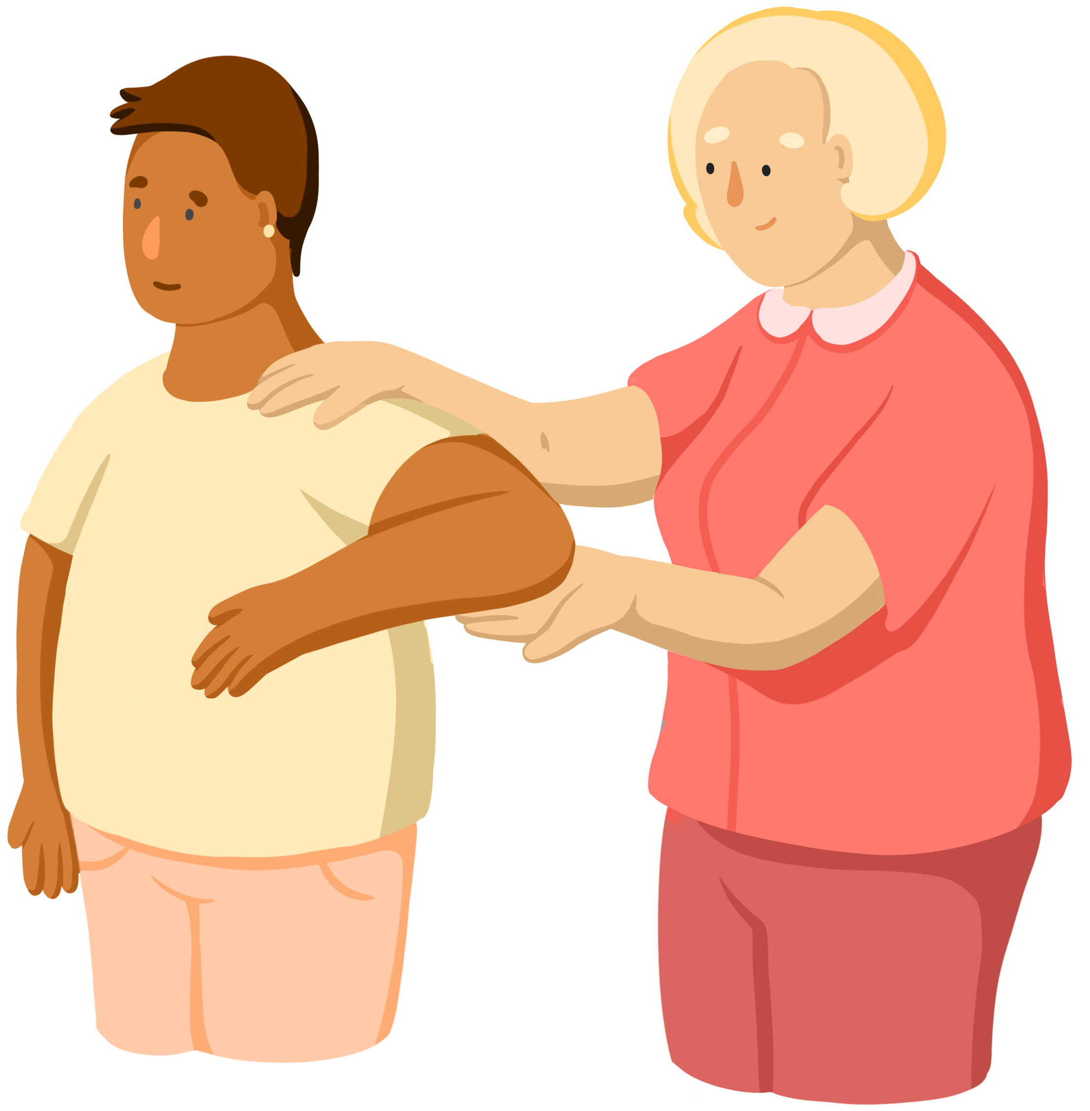
Physiatrists (fiz-EYE-uh-trists) are doctors that focus on how the body moves. They pay attention to how your body works, not just where it hurts. They are can diagnose and treat pain or movement problems.
Your physiatrist may check how well you can move by asking you to do simple tasks. They may also use nerve tests to take a closer look at your nerves and muscles.
You may see a physiatrist more than once at the clinic because they are involved in many stages of your treatment.
Surgeons
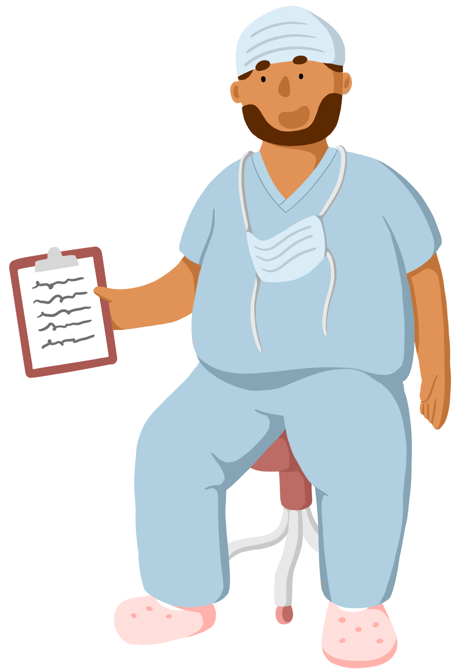
Surgeons (SUR-jun) are doctors that perform operations to help your body heal faster. At the clinic, a surgeon may talk to you if your care team thinks surgery might help you recover better.
There are different kinds of nerve surgeries possible. Your surgeon will explain what the surgery involves, how long it will take, and what to expect during recovery.
After surgery, surgeons will continue to check in on your healing and may suggest other treatments to help you recover.
Physical Therapists
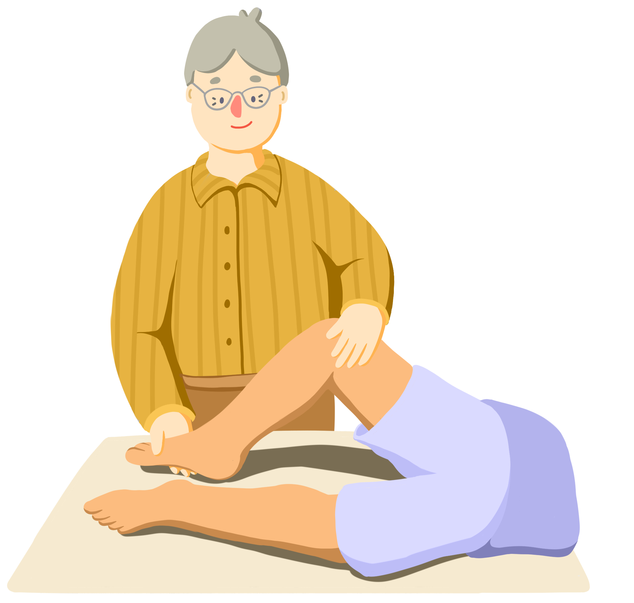
A physical therapist (FIZ-eh-kul THER-uh-pist) or PT will find ways to reduce your pain and improve your ability to move. They might show you exercises or stretches to do at the clinic or at home to help you regain movement in your arm or leg.
You might see a PT several times depending on your recovery plan. They can help keep track of how you're doing and adjust your exercises as you get stronger.
Occupational Therapists
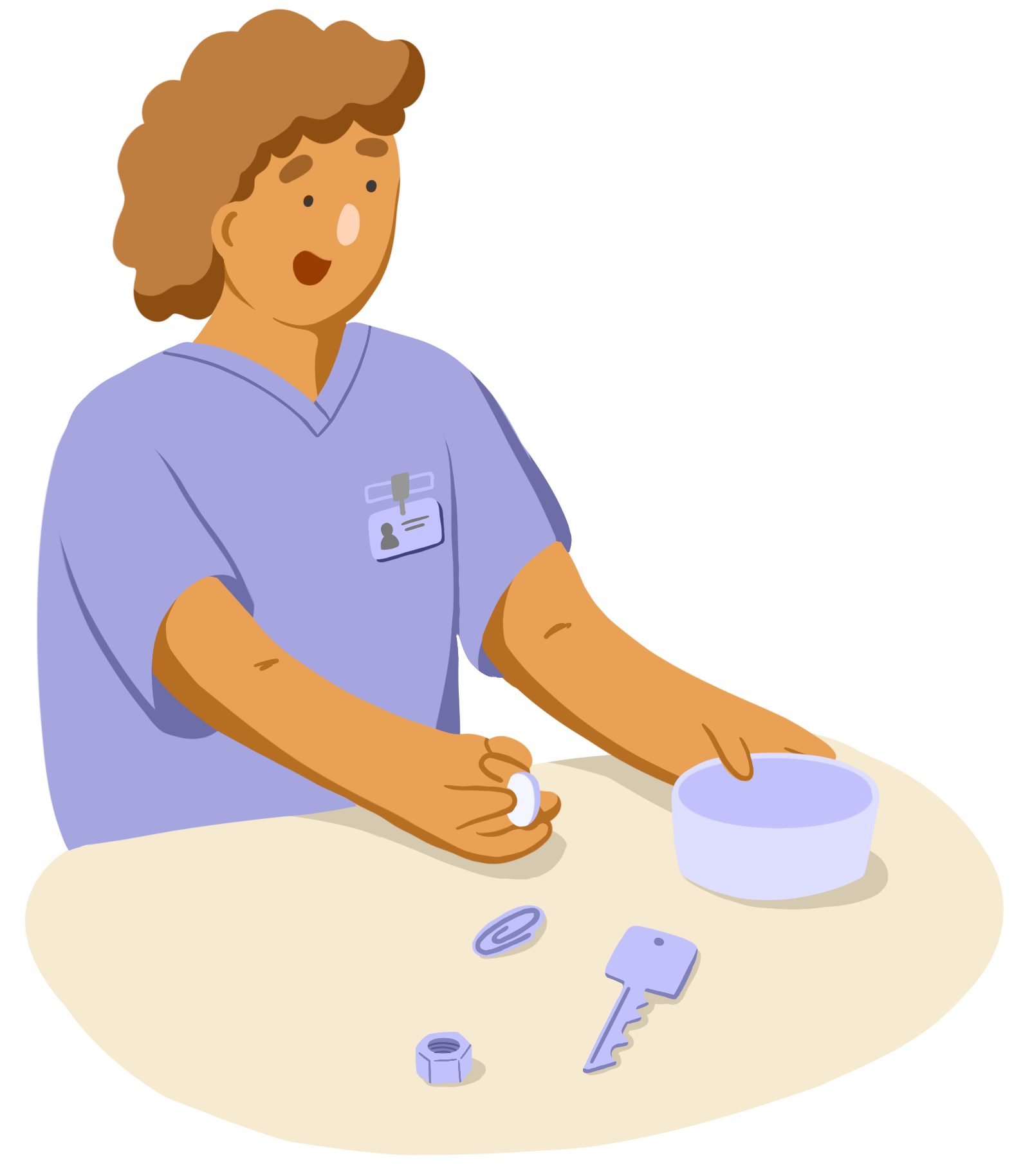
An occupational therapist (ohk-you-PAY-shun-uhl THER-uh-pist) or OT will help you adjust to life with peripheral nerve injury. If daily tasks like picking up small objects feel harder after your injury, they can help you find new ways to do them.
OTs may suggest tools like braces or splints to support your injured arm or leg. These can help make daily tasks easier while keeping you safe.
You may see an OT several times depending on your recovery plan. Over time, they can help you adjust to changes in your life and find new strategies or other helpful tools.
All of these care providers work together to make sure you're getting the support you need at each stage of recovery.
Recovery can be difficult and frustrating, especially when it has impacted your daily life or how you see yourself. If you are feeling overwhelmed or concerned, reach out to your care team. They are always there to provide support and help you during your PNI journey.
Diagnostic Testing
Diagnostic tests can help doctors find the cause of your symptoms and understand how serious the nerve injury is. This information helps them choose the best treatment plan for you.
Medical History and Physical Exam
Before conducting any tests, the doctor will ask you questions about your injury. They will also look at your medical history to see if there are any health problems that could impact treatment and recovery.
The doctor will also check the injured area. You might be asked to do some simple movements to figure out which muscles and nerves have been affected.
Medical Imaging
To get a closer look at the injury, you may be asked to do some imaging exams such as:
- X-rays
- CT scans
- MRIs
- Ultrasounds
X-rays and CT scans both radiation that has a small risk of harming tissue. However, the risk of developing cancer from this exposure is quite low. X-rays and CT scans are incredibly helpful tools for doctors to diagnose conditions to get you the care you need.
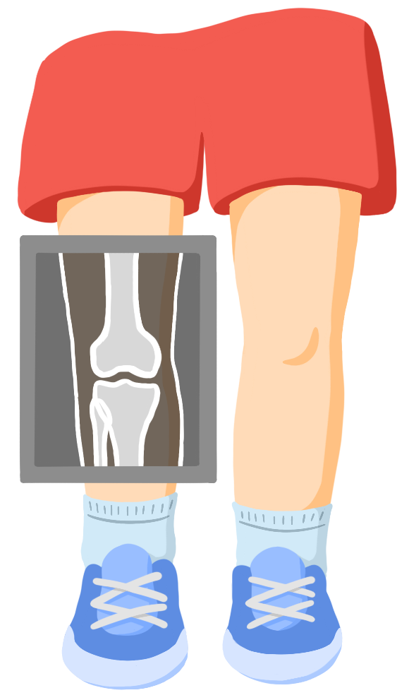
X-ray
X-rays show images of structures inside the body, such as your bones and organs.
During an x-ray, radiation is passed through your body. Tissues that absorb x-rays easily, such as bone, will appear whiter on the x-ray. The final image is created from the different amounts of radiation that reach the detector.
X-rays can help doctors see major bone injuries that might be around the nerve.
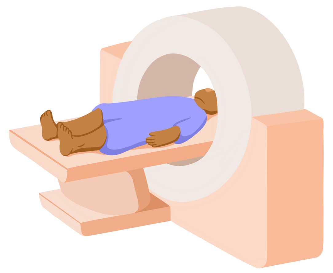
CT Scan
CT scans use stacked x-rays to create detailed, cross-sectional images of the body. They are also good at showing hard structures such as bone, but are more detailed than x-rays.
During a CT scan, you lie on a bed that moves through a CT machine. As you move through the machine, x-rays will pass through your body. With new machines, CT scans can be done quickly in 15 minutes or less.
Doctors use CT scans to see bone injuries in detail and get a better look at the structures around the nerve.
MRI
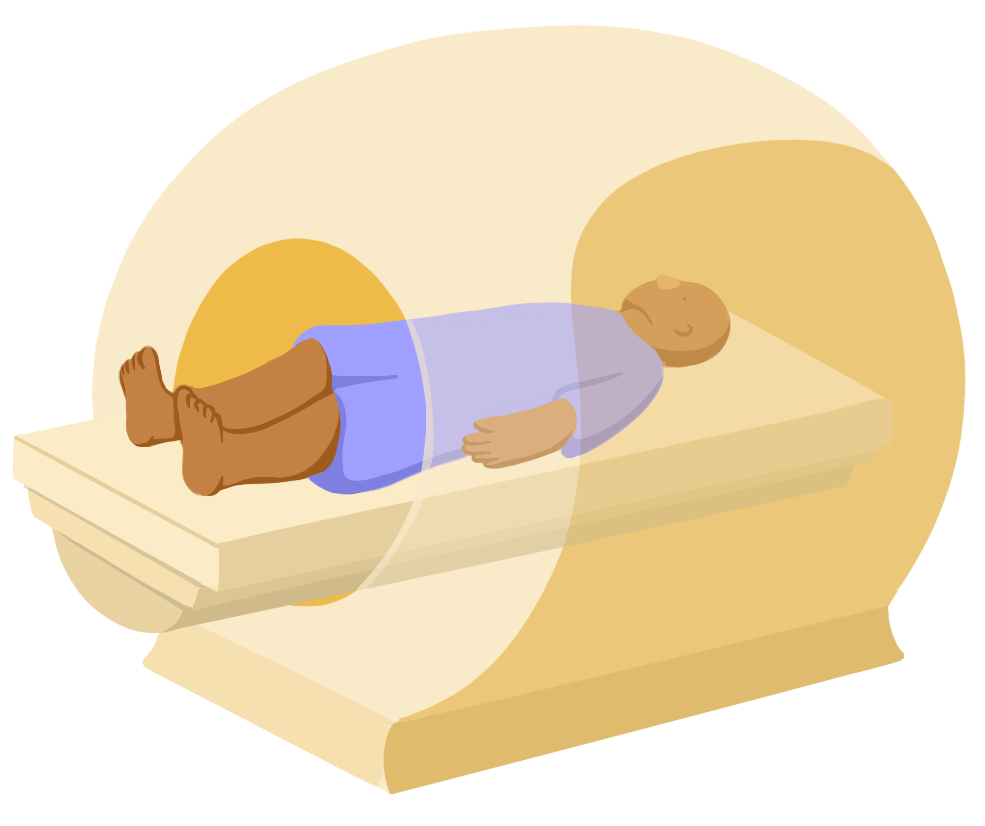
Instead of radiation, magnetic resonance imaging (MRI) uses radio waves and magnetic fields to create cross-sectional images of your body.
They are good at getting detailed pictures of soft tissues, such as nerves and muscles.
During an MRI, you lie on a bed that moves through an MRI machine. This machine acts like a giant magnet that sends radio waves through your body. This scan can take up to an hour to complete depending on how much of your body needs to be scanned.
Doctors use MRIs to get more detailed images of nerves and the injury site.
Ultrasound
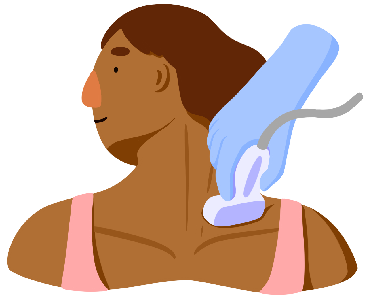
Ultrasounds help doctors see nerves and the surrounding area in real-time and during movement.
During an ultrasound, a device called a transducer is placed over the skin. The transducer passes sound waves through the body. Different tissues will reflect the sound waves in different ways to show the location and shape of bones, muscles, nerves, and more.
There are no known risks for getting an ultrasound, but sound waves don’t travel as well through the body, so the image may not be as clear.
Nerve Tests
Doctors can look at the electrical signals passing from nerves to muscles. If the signals are unusual, it might mean that there is a problem with the path between the nerve and muscle. The size and speed of the signal can help identify the severity and type of nerve damage that has occurred.

Click through the different tabs to explore the different nerve tests.
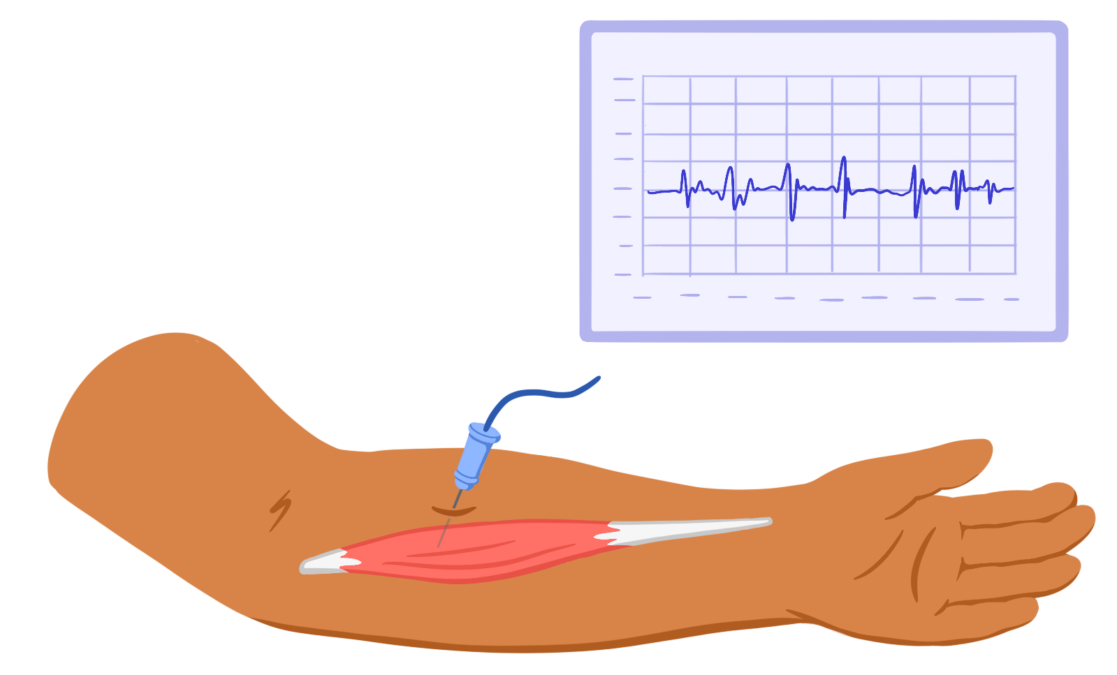
Needle Electromyography (EMG)
Electromyography (ee-LEK-troh-my-OG-ruh-fee) or EMG is a nerve test that measures the electrical activity of muscles and nerves.
EMGs will use needle electrodes, which are special, small needles that measure electrical signals. During the test, the needle electrode is inserted through the skin. You may be asked to stay still or gently contract your muscles.
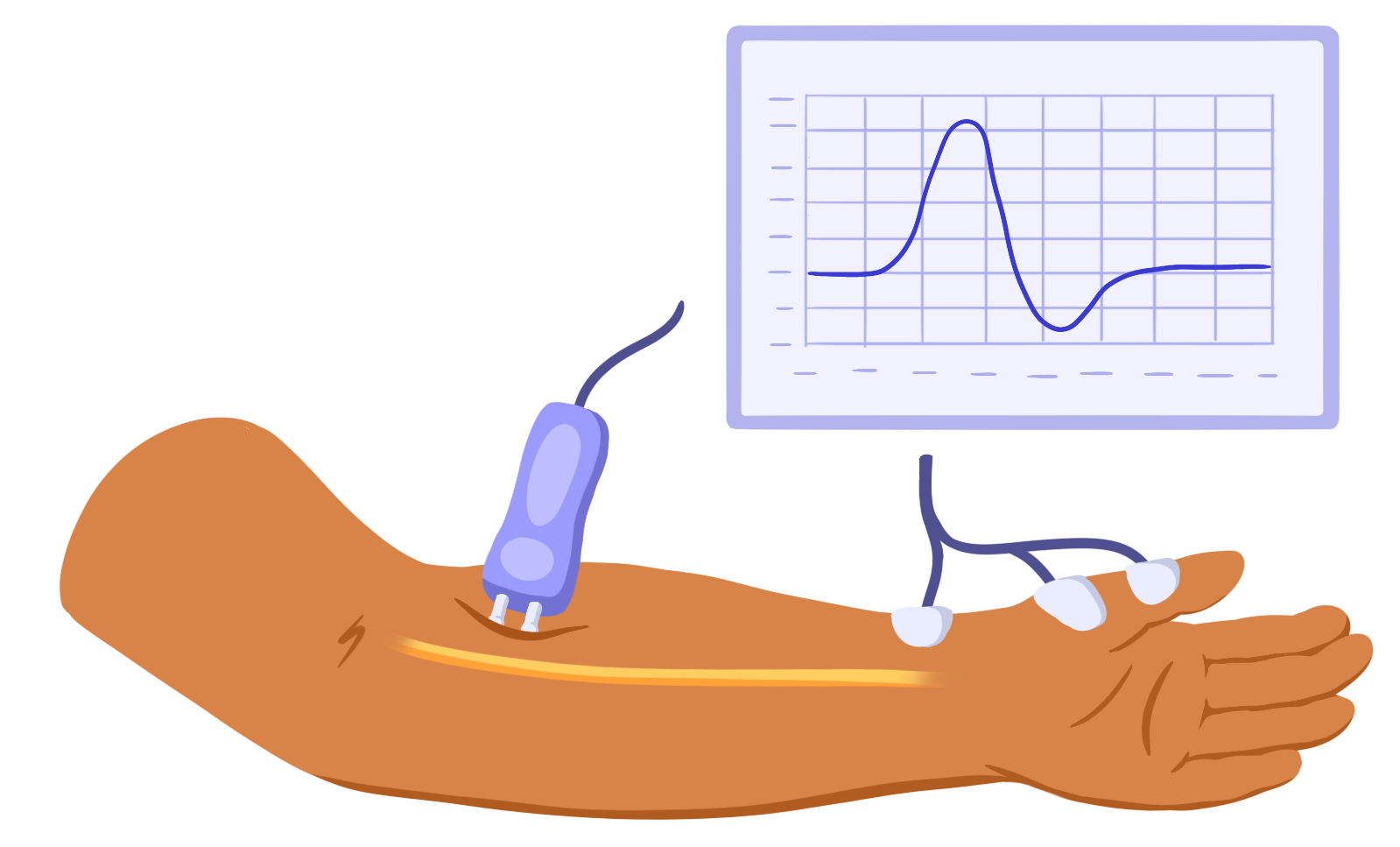
Nerve Conduction Test
A nerve conduction test measures the speed and strength of the electrical signals passing in a nerve.
Nerve conduction studies use surface electrodes. These can measure electrical signals through the skin. Surface electrodes are placed on your skin at different points. A brief, mild electric pulse will be sent through one electrode to the other.
While EMG looks at the path of electrical signals from the nerve to the muscle, a nerve conduction study looks at the path of electrical signals along the nerve itself.